6 December 2024 : MRI Symposium : Focus on the new technologies
The next MRI Day is scheduled for Friday 6 December at Genopolys. We would like to thank the speakers who have agreed to share their science and illustrate their experience with MRI. The focus will be on the latest technologies installed on MRI. You will find all the information on the poster.
Don't forget to register before 31 October here.

October 2024 : New confocal microscope in the new MRI-PHIV-DIADE facility
A new facility has been set up at the IRD within the UMR DIADE. It is run by Carole Gauron, who manages an AxioZoom macroscope and a new Leica confocal microscope, a Stellaris 8. All the details are on the dedicated webpage.
Thanks to contact This email address is being protected from spambots. You need JavaScript enabled to view it. for more informations and to discuss your projects
October 2024 : MRI-ISEM at the Fête de la Science
Renaud Lebrun, manager of the MRI-ISEM X-Ray tomography facility and his colleague Mavea Orliac will be welcoming you on saturday 5 october for the Fête de la science. They'll be telling you all about the evolution of cetaceans. Don't hesitate to go and see them

October 2024 : MRI present to the first meeting of BioCampus experts
The MRI facility had the opportunity to present its activities and expertise at the morning session organised by the UAR BioCampus on the theme of ‘Pre-clinical models and alternatives’. The main MRI help in this area is to characterise and screen the models proposed by the RAM, ZEFIX, POM and DROSO facilities.
For more information, please contact us at This email address is being protected from spambots. You need JavaScript enabled to view it.
September 2024 : MRI present at Phot'Aubrac
As a follow-up to the 2023 photo exhibition ‘La vie est belle’, MRI exhibited a selection of photos at the Phot'Aubrac. festival. As at the Jardin des Plantes last year, the exhibition was a great success with the general public and we enjoyed explaining our professions. An experience worth repeating!

September 2024 : the MRI-EM4Bio platform present to the CRYO-EMontpellier symposium
Laurence Berry, head of the new MRI-EM4bio platform, had the opportunity to present her technological solutions during the day dedicated to CRYO-EM. Feel free to see what was on offer on the dedicated webpage.
Please contact This email address is being protected from spambots. You need JavaScript enabled to view it. for more information or to discuss your projects.
June 2024 : New confocal microscope in the MRI-INM facility
The MRI-INM facility offers access to a new Leica confocal microscope. This is a Stellaris 5 equipped with a white laser, hybrid detectors and 6 objectives, including 2 long-distance objectives. Full description of this machine on the dedicated webpage
Please contact This email address is being protected from spambots. You need JavaScript enabled to view it. for more information or to discuss uour projects
June 2024 : MRI present to the Occitanie Réseau Imagerie meeting in Perpignan
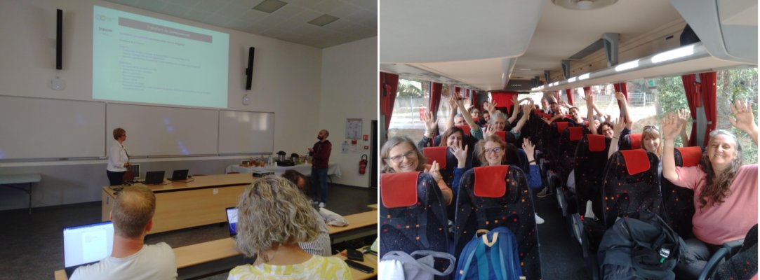
Like every year, MRI took part in the Occitanie imaging network day. This year, an expedition was organised to join our colleagues from Toulouse, Perpignan and Banyuls-sur-mer in Perpignan. It was a day rich in scientific and professional exchanges! See you next year.

May 2024 : MRI present to the ANF ApiPhot - Assises RTmFm
Few MRI engineers attended the APiPhot training course organised to mark the 20th anniversary of our national RTmFm network. Orestis Faklaris and Julio Mateos-Langerak presented the work they are involved in as part of RTmFM or FBI working groups. MRI is also present in the Microscopoly created by the RTmfm!

April 2024 : MRI is looking for an engineer in Flow cytometry and Photonic microscopy
We are looking for an engineer who will spend his/her working time between 2 assignments within the MRI facility: facility manager for the IGH cytometry facility and work as an cytometry engineer and microscopy engineer on the IGH microscopy facility. The fixed-term contract starts on 15 June 2024 for one year and is renewable. The profile is available on the CNRS job portal:ici
Please, feel free to contact This email address is being protected from spambots. You need JavaScript enabled to view it. or This email address is being protected from spambots. You need JavaScript enabled to view it. for more informations.
March 2024 : MRI is looking for a technician for Electron Microscopy
We are looking for a technician dedicated to the sample preparation in electron microscopy. The fixed-term contract will run from as soon as possible until 31/12/2025. This person will work on the new MRI-EM4Bio facility recently created in building 24 on the Triolet Campus of the University of Montpellier. The profile is here.
2024 : Our training shedule for the year:
MRI has scheduled all its training activities (BioCampus workshops). These courses are given in french. The dates are as follows:
Microscopie à épi-fluorescence et microscopie confocale : de la base à la pratique : 4-7 march
Cytométrie en flux: de la préparation des échantillons à l’analyse des données: 19-20 march
Programmation des macros avec Image J: 26-28 march
Les bases du logiciel Image J : 25,26 et 29 april
L’apprentissage automatique (machine learning)appliqué à l’analyse d’images biologiques : 4-6 june
Imagerie avancée : Haute résolution en microscopie photonique : 26-26 june
Analyse d’images 3D : 8-10 october
Formation FlowJo niveau débutant : 7 november
January 2024 : Best wishes :
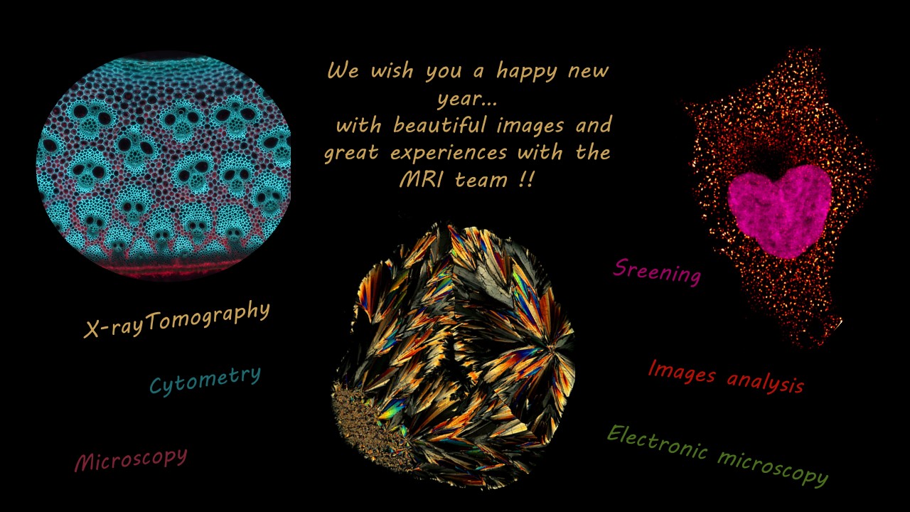
MRI wishes you all a very happy new year 2024. As every year, MRI staff will be on hand to provide you with the best possible support for your research projects.

9-17 november 2023 : MRI at Mifobio :
6 MRI engineer and the MRI scientific director attended the latest edition of the Mifobio 2023 theme school on the Giens peninsula. An ideal opportunity to learn about the latest technological advances and an excellent way to share our experience through workshops and round tables.

october 2023 : MRI video online! :
As part of its 20th anniversary celebrations, MRI has produced a new video of the facility, looking backwards and forwards.
In preview!
17 - 19 octobre 2023 : Symposium "Imagine Life : the Future" :
As part of its 20th anniversary celebrations, MRI organised a symposium at the Corum in Montpellier. This 2.5-day symposium aims to examine how future developments in light/electron microscopy and image analysis could further transform research in animal and plant biology at multiple scales, from molecules to tissues. The symposium will include 7 thematic sessions (image analysis, cancer biology, plant biology, 3D genome architecture and transcription, neurosciences, cell and developmental biology, infection and immunity).
29 september - 31 october 2023 : Photo exhibition "La vie est belle"
MRI is organising a photo exhibition of 44 large-format science photos entitled "La Vie est belle!" as part of its 20th anniversary celebrations. This exhibition is open at the Montpellier Jardin des Plantes (Blvd Henri IV) until 31 October. This exhibition combines 32 scientific photos taken on the MRI platform and communicated by its users and 12 ambient photos, which show the process of capturing these images.
Entrance to the exhibition is free and the venue magical. Don't miss the exhibition and come along with your family or friends: you don't have to be a scientist to enjoy it!

21 september 2023 : Seminar by Laurence Berry about the new MRI-EM4Bio electron microscopy facility
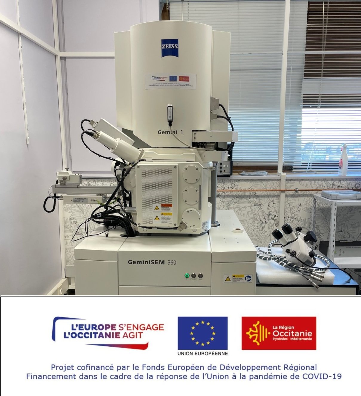
On the occasion of the creation of an electron microscopy service on MRI, we are pleased to announce a seminar to be held on 21 September at 9.45am in room sc36.09 of the Triolet Campus (new white building at the entrance to the University). Laurence Berry, who is in charge of the facility, will explain the range of technologies and applications we can offer you, based on the use of the microscope recently installed in building 24, the Zeiss Gemini 360 and the volutome. This microscope was funded as part of the European ReactEU program.
A dedicated webpage is currently under contruction.


