R
- Details
- open source software
R is a free software environment for statistical computing and graphics.
- Available at MRI-CRBM
- https://www.r-project.org/
The facility provides access to proprietary analysis software, open source software and tools developed in house. We also provide access to deep-learning solutions including the training on your own data on our machines.
R is a free software environment for statistical computing and graphics.
Software dedicated to FLIM image analysis. Allows, among other things, calculations of fluorescence lifetime, amplitudes, and intensities.
Automatic detection and segmentation of cells and nuclei using star-convex polygons.
Use dl4mic to train and apply StarDist.
See symphotime-64-fluorescence-lifetime-imaging-and-correlation-software
UNet is a deep-learning method for pixel classification. It can be trained to segment different kinds of objects in images. The first half of the U-Net architecture is a downsampling convolutional neural network which acts as a feature extractor from input images. The other half upsamples these results and restores an image by combining results from downsampling with the upsampled images. Use dl4mic to train and apply the UNet.
Moving seamlessly among restoration, visualization and quantitation, Volocity software is designed for true 3D analysis of fluorescence images. View your cells from every angle. Measure shapes, volumes and distances. Relate cellular structure to function with exceptional precision and speed. Compare samples and identify trends. Produce publication-ready tables and charts. Take your image analysis to a new dimension with true 3D image analysis.
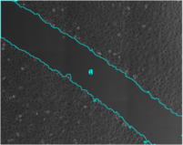
The tool can be used to analyze scratch assays. It measures the area of a wound in a cellular tissue on a stack of images representing a time-series.
Download and further informations: Wound Healing Tool on github
Image analysis software. Allows to work with 3D images in the ZEISS format. It's possible to analyse FRAP experiments and if needed to process the (fast) AiryScan data.
A number of image processing and analysis software packages are available on our analysis computers. Please contact us if you need help with on of the software packages.
A number of open source image analysis software packages are available on our computers. Other open source packages can be installed on demand. Most of our computers are equipped for 3D/4D processing and also for training and applying deep learning solutions. If you need help with a open source image analysis software please contact us.
Analysis tools and extensions developed at MRI. You can find the complete list of publicly available tools we developed in our github. Please contact us if you have an unsolved image analysis problem. We can either point you to already existing solutions of develop a custom tool for you.
We developed a tool dl4mic, that allows are users to train and run deep learning solutions from a graphical user interface integrated into FIJI/ImageJ. Currently 4 network-models are available within the tool: UNet, Dense-UNet, Noise2Void and Stardist. The tool allows to quickly integrate further models. For cell-segmentation we propose Cellpose. Cellpose is already trained with a large number of cell images and can often be used without further training. Our version of cellpose allows to run batch processing from within the graphical user interface.